Suggested protocol for dynamic mri of pelvic floor dysfunction.
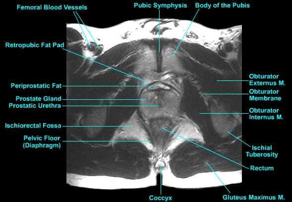
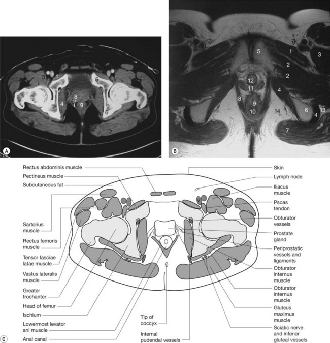
Male pelvic floor mri.
Use the mouse scroll wheel to move the images up and down alternatively use the tiny arrows on both side of the image to move the images on both side of the image to move the images.
Magnetic resonance imaging mri dynamic pelvic floor.
The male pelvic floor has sex specific differences in anatomy and pathophysiologic disorders.
A pelvic mri scan uses magnets and radio waves to help your doctor see the bones organs blood vessels and other tissues in your pelvic region the area between your hips that holds your.
Prostate bladder genital organs rectum many thanks to samuel merigeaud md for his medical contribution.
Many variations in the techniques described below exist.
Mr defecating proctography is a dynamic study for evaluation of the pelvic floor and pelvic organ prolapse.
Overflow incontinence is less common in women than in men.
Functional disorders of the pelvic floor such as pelvic organ prolapse and defecatory dysfunction represent a common health problem especially in women it is estimated that more than 15 of multiparous women 1 are affected by some sort of pelvic disorder and that 10 20 of patients seek medical care in gastrointestinal clinics for evacuation dysfunction 2.
View larger version table 1.
Dynamic pelvic floor magnetic resonance imaging mri is a noninvasive test that uses a powerful magnetic field radio waves and a computer to produce detailed pictures of the pelvic floor a network of muscles that stretches between the pubic bone and spine and the abdominal organs it supports.
Zonal anatomy adapted for sector map adapated frompi rads v2 acr.
Anatomy of the male pelvis on mr imaging.
There are four phases of evaluation.
The prevalence of pelvic floor disorders is much lower in men than in women and because of this the majority of the published literature pertaining to mri of the pelvic floor is oriented toward evaluation of the female pelvic floor.
Pelvic floor failure is a common disorder that affects 23 7 of women in the united states with a prevalence of 9 7 49 7 that increases with age one in nine women will undergo an invasive procedure for treatment of urinary incontinence or pelvic organ prolapse with 30 requiring additional surgery for symptom recurrence by 80 years of age.
Interpretation of mri findings.
A suggested protocol for mri of pelvic floor dysfunction is summarized in table 1.
This mri male pelvis axial cross sectional anatomy tool is absolutely free to use.
Despite these differences static and dynamic mri features of these disorders specifically gastrointestinal disorders are similar in both sexes.